
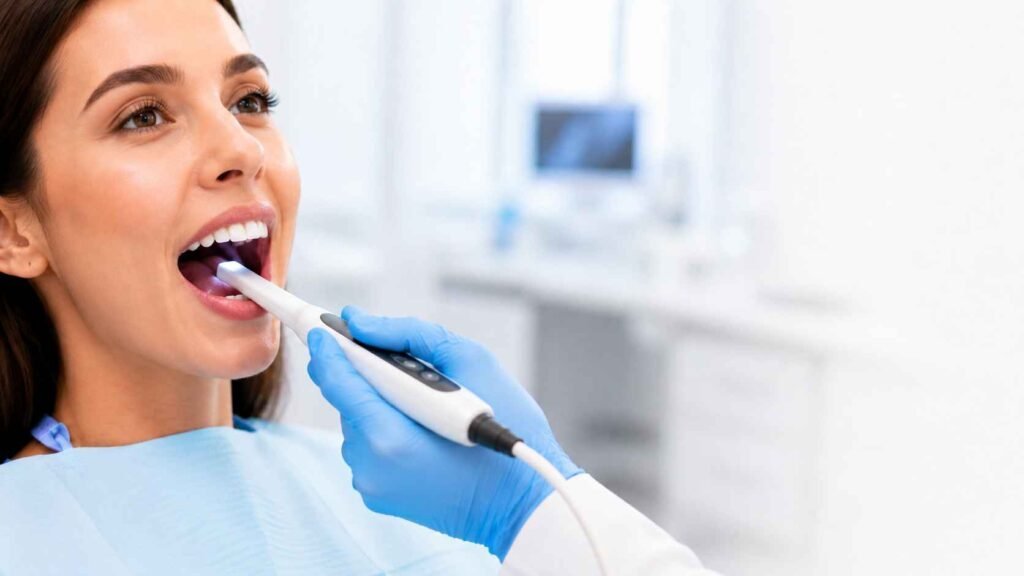
What does an intraoral camera show?
Many problems in the mouth cause no pain and are hard to spot in a hand mirror. On the screen you can see a chipped edge or a fine crack for yourself. The wand itself is about the size of a thick marker.
- cracks, chips and worn biting edges
- the edges of older fillings and crowns
- plaque, tartar and stains
- gum colour and swelling along the gumline
- spots on a tooth surface that need a second look
Finding decay at its earliest stage has its own tools, and our early cavity detection page explains them.
How does the camera work?
The camera works like a tiny video camera with its own LED light, which reaches back teeth that an overhead lamp misses. The wand moves slowly along your teeth while the picture plays on the monitor.
Taking photos needs no numbing. You feel the wand resting against your teeth and gums, much like a dental mirror. Tell the team if you have a strong gag reflex or a sore jaw, and ask for a pause whenever you need one.
The wand touches the inside of your mouth, so Ontario's infection prevention and control standard for dental offices covers it. Under that RCDSO standard, a camera is either cleaned and heat-sterilized between patients or covered with a barrier. After the barrier comes off, the camera is checked. If it is soiled, it is cleaned and then disinfected.
Want to see your own teeth on the screen? Ask at your next checkup.
What can't the camera show you?
The camera shows surfaces only. It cannot see inside a tooth, between teeth where they touch, under the gums, around the roots or into the jawbone. Decay that starts between teeth, infection at a root tip and bone loss stay hidden from it.
Those areas need dental X-rays, and the camera does not replace them. Health Canada's Safety Code 30 says dental X-rays follow a clinical exam and a review of your history. They are not a routine screen. So the dentist decides whether you need any after your history and exam.
A photo is one part of the exam. It cannot by itself confirm or rule out a cavity or gum disease. Saliva and lighting change how a spot looks on screen, so the dentist reads the photo together with the rest of your exam.
Where do your photos go, and who can see them?
Photos the dentist saves become part of your digital dental record at My City Dental. Photos of the same tooth over time let the dentist compare whether a crack or the gumline has changed. The clinic can send them to another dental office involved in your care, to your dental insurer or to a dental lab making a crown.
In Ontario, your dental record is personal health information under the Personal Health Information Protection Act (PHIPA). That law lets you give or refuse consent to how your information is shared, with some exceptions.
Direct insurance billing. Canadian Dental Care Plan (CDCP) accepted. CDCP coverage depends on the service and your eligibility, and some services need preauthorization.
Common questions
Why would a dentist use an intraoral camera?
A dentist uses the camera to judge small surface details and to show them to you. A hairline crack or a lifting filling edge is easier to assess when it fills a screen. Seeing the problem yourself makes the reason for a suggested treatment easier to follow.
Does the camera use radiation like an X-ray?
No. The camera takes ordinary digital photos using the light from a small LED on its tip, so it gives off no X-rays. That is also why it cannot see through enamel or gum tissue. X-rays look inside a tooth and into the bone, and the dentist decides whether you need any after your history and exam.
Is there a separate charge for intraoral photos?
Ask the team before your visit whether intraoral photos carry a separate fee. Your own cost comes in a written estimate after an exam. Insurance plans set their own rules for photos, so check with your plan as well. CDCP patients can ask the team what applies to them before any treatment starts.
When the dentist may use the camera
The camera helps at many kinds of visits, wherever an area is hard to see clearly.
- During a routine checkup
- To check small cracks or worn tooth edges
- Before recommending a filling, crown or other treatment
- To watch for changes along the gumline
- To look around older fillings and crowns
- When a tooth feels sensitive or sore
- To record visible changes in a tooth over time
How intraoral photos help you
Clear photos make it easier to follow what the dentist found during your exam.
- Close-up views of your teeth and gums
- A clearer picture of why a treatment is suggested
- Small problems you can see for yourself
- Before and after photos of treated teeth, for comparison
- An easier conversation with the dentist
- More information before you choose a treatment